Sharp Laboratory : Structural Mass Spectrometry

​Dr. Sharp’s group is active in the development and application of new mass spectrometry-based technologies for studying the structure-function relationships of proteins and carbohydrates of biomedical interest. Current applications focus on the study of interactions between neutralizing antibodies and the glycoprotein coat of HIV; characterization of chemokines and the structural factors mediating their oligomerization and function; and the identification of glycosaminoglycan structures with potential biomedical applications for a wide variety of fields including anti-microbial agents, anti-cancer therapies, and anti-inflammatory therapies. 

Overview

Recent Selected Publications

Farrag M, Aljuhani R, Benicky J, Al Ahmed H, Misra SK, Mishra SK, Sharp JS, Doerksen RJ, Goldman R, Pomin VH. Heparan-6-O-endosulfatase 2, a cancer-related proteoglycan enzyme, is effectively inhibited by a specific sea cucumber fucosylated glycosaminoglycan. Glycobiology. 2025 Apr 23;35(6):cwaf025. doi: 10.1093/glycob/cwaf025. PMID: 40302034; 

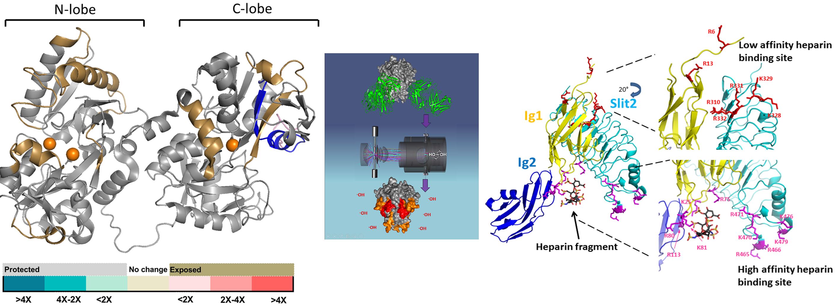
Heparan-6-O-endosulfatase 2 (Sulf-2) is a proteoglycan enzyme that modifies sulfation of heparan sulfate proteoglycans. Dysregulation of Sulf-2 is associated with various pathological conditions, including cancer, which makes Sulf-2 a potential therapeutic target. Despite the key pathophysiological roles of Sulf-2, inhibitors remain insufficiently developed. In previous work, a fucosylated chondroitin sulfate from the sea cucumber Holothuria floridana (HfFucCS) exhibited potent Sulf-2 inhibition. This study investigates the structural basis of HfFucCS-mediated Sulf-2 inhibition, examines the binding profile of HfFucCS to Sulf-2, and explores the mode of inhibition. Additionally, a structurally diverse library of sulfated poly/oligosaccharides, including common glycosaminoglycans and unique marine sulfated glycans, was screened for Sulf-2 inhibition. Results from a high-throughput arylsulfatase assay and specific 6-O-desulfation assay have proved that HfFucCS is the most potent among the tested sulfated glycans, likely due to the presence of the unique 3,4-disulfated fucose structural motif. HfFucCS demonstrated non-competitive inhibition, and inhibitory analysis of its low-molecular-weight fragments suggests a minimum length of ~7.5 kDa for effective inhibition. Surface plasmon resonance analyses revealed that Sulf-2 binds to surface heparin with high affinity (KD of 0.817 nM). HfFucCS and its derivatives effectively disrupt this interaction. Results from mass spectrometry-hydroxyl radical protein footprinting and repulsive scaling replica exchange molecular dynamics indicate similarities in the binding of heparin and HfFucCS oligosaccharides to both the catalytic and hydrophilic domains of Sulf-2. These findings reveal the unique inhibitory properties of a structurally distinct marine glycosaminoglycan, supporting its further investigation as a selective and effective inhibitor for Sulf-2-associated cancer events.

----------------------------------------------------------------------------------------------------------------

Bohnsack RN, Misra SK, Liu J, Ishihara-Aoki M, Pereckas M, Aoki K, Ren G, Sharp JS, Dahms NM. Lysosomal enzyme binding to the cation-independent mannose 6-phosphate receptor is regulated allosterically by insulin-like growth factor 2. Sci Rep. 2024 Nov 6;14(1):26875. doi: 10.1038/s41598-024-75300-9. PMID: 39505925.

The cation-independent mannose 6-phosphate receptor (CI-MPR) is clinically significant in the treatment of patients with lysosomal storage diseases because it functions in the biogenesis of lysosomes by transporting mannose 6-phosphate (M6P)-containing lysosomal enzymes to endosomal compartments. CI-MPR is multifunctional and modulates embryonic growth and fetal size by downregulating circulating levels of the peptide hormone insulin-like growth factor 2 (IGF2). The extracellular region of CI-MPR comprises 15 homologous domains with binding sites for M6P-containing ligands located in domains 3, 5, 9, and 15, whereas IGF2 interacts with residues in domain 11. How a particular ligand affects the receptor's conformation or its ability to bind other ligands remains poorly understood. To address these questions, we purified a soluble form of the receptor from newborn calf serum, carried out glycoproteomics to define the N-glycans at its 19 potential glycosylation sites, probed its ability to bind lysosomal enzymes in the presence and absence of IGF2 using surface plasmon resonance, and assessed its conformation in the presence and absence of IGF2 by negative-staining electron microscopy and hydroxyl radical protein footprinting studies. Together, our findings support the hypothesis that IGF2 acts as an allosteric inhibitor of lysosomal enzyme binding by inducing global conformational changes of CI-MPR.

-----------------------------------------------------------------------------------------------------------------

Tobin L, Misra SK, Luo H, Jones LM, Sharp JS. Radical Protein Footprinting in Mammalian Whole Blood. bioRxiv [Preprint]. 2024 Sep 29:2024.09.29.615683. doi: 10.1101/2024.09.29.615683. PMID: 39386581.

Hydroxyl Radical Protein Footprinting (HRPF) is a powerful method to probe the solvent-accessible surface area of proteins. It is mostly used to study the higher-order structure of proteins, as well as protein-protein and protein-carbohydrate interactions. Hydroxyl radicals are generated by the photolysis of hydrogen peroxide and these radicals modify the surface amino acids. Bottom-up proteomics is then applied and peptide oxidation is calculated and correlated with solvent accessibility. It is mainly performed in vitro; however, it has been recently used in living systems, including live cells, live nematodes, and 3D cell cultures. Mammalian tissues are still out of reach as they absorb UV strongly, hindering radical generation. Here, we describe the first example of RPF in mammalian stabilized whole blood. Using photoactivation of persulfate with a commercially available FOX Photolysis System modified for sample handling and inline mixing, we demonstrate the first labeling of proteins in whole blood. We demonstrate that the RPF protocol does not alter the blood cell gross morphology outside of a moderate hypertonicity equivalent to sodium chloride exposure prior to labeling. We detail an improved quenching protocol to limit background labeling in persulfate RPF. We describe the labeling of the top ten most abundant proteins in the blood. We demonstrate the equivalence of ex vivo labeling in whole blood with labeling of the same structure in vitro using hemoglobin as a test system. Overall, these results now open the possibility of performing RPF-based structural proteomics in pre-clinical models and using readily available clinical samples.