Research Cores

The Center for Nano-Bio Interactions (CNBI) at the University of Mississippi is a thriving center for pioneering advancements in biomaterials and nanobiotechnology research. CNBI's two core suites offer cutting-edge capabilities in materials characterization and biological analysis. The Biomaterials Characterization Suite is outfitted with a range of instruments for soft matter characterization, including thermal light scattering, and mechanical analysis. The Biomolecular Analysis Suite offers capabilities ranging from traditional molecular biology (PCR, SDS-PAGE, Gel Imaging, etc.) to high-resolution biomolecular techniques such as flow cytometry, flow-assisted cell sorting, confocal microscopy, and single cell analysis. Available through the Biomolecular Analysis Suite are also IVIS imaging and a Seahorse for metabolic analysis. With a commitment to cultivate a culture of inclusivity, knowledge sharing, providing training and support, the core extends its resources to a diverse range of individuals, spanning from faculty to ambitious undergraduate researchers alike, throughout their scientific journey. The CNBI cores serve as a catalyst for interdisciplinary collaboration, technological innovation, and scientific excellence, paving the way for a brighter future in biomedical research in the state of Mississippi and beyond.


Book an Instrument

Biomaterials Characterization Suite

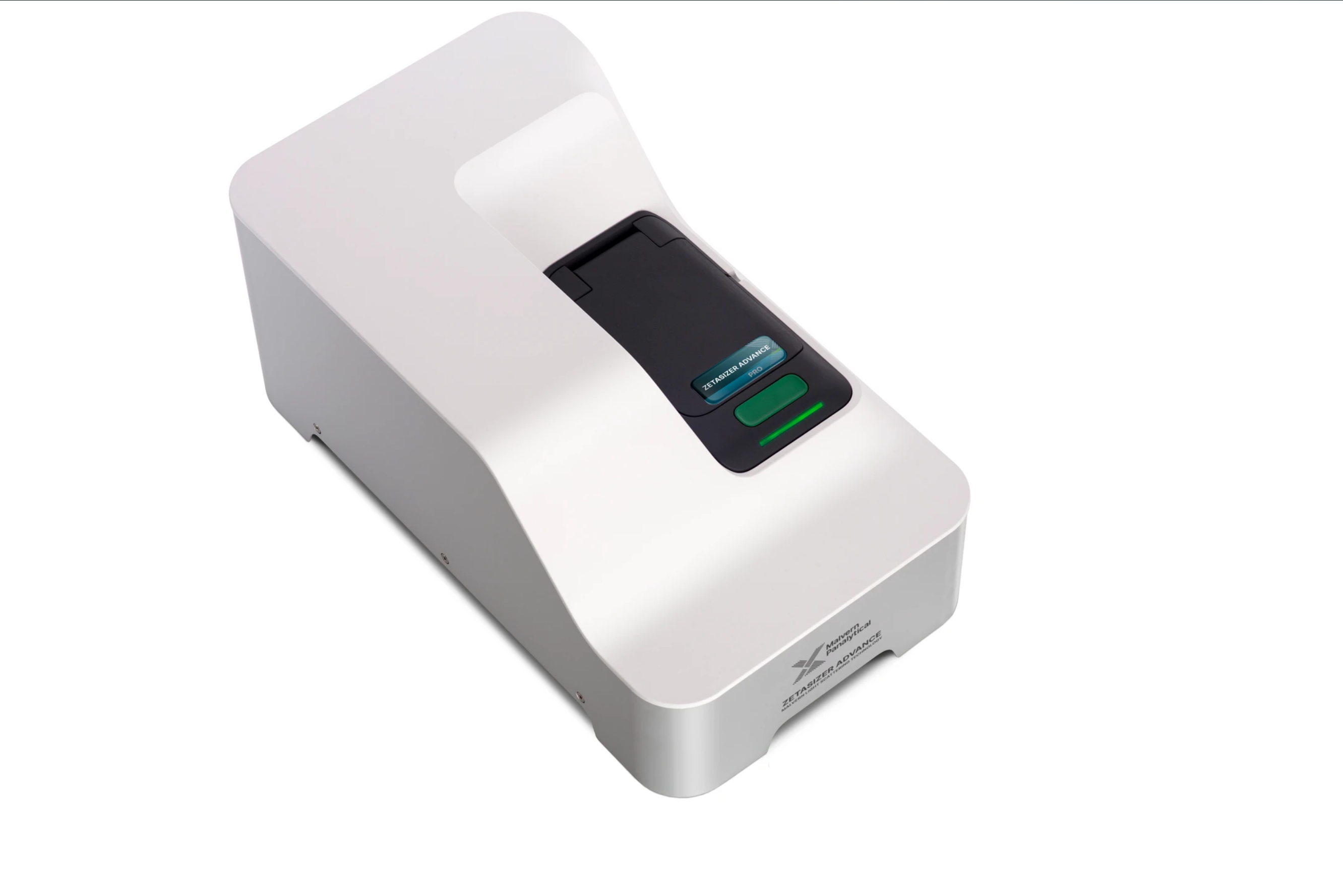
Malvern Zetasizer Pro

The Malvern Zetasizer Pro offers wide dynamic concentration capability and high sensitivity due to the Non-Invasive Back Scatter (NIBS) optical design. Measurement of particle size, molecular size, electrophoretic mobility, zeta potential and molecular weight can all be performed with ease using the instrument. 

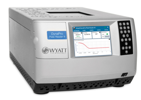
DynaPro Plate Reader III

The DynaPro DLS/SLS Plate Reader is a high-performance instrument for dynamic and static light scattering analysis, designed to characterize proteins and nanoparticles. It features both DLS for measuring particle size and SLS for determining molecular weight, all within a high-throughput microplate format. The system employs advanced optics for precise measurements and comes with intuitive software for streamlined data acquisition and analysis, making it ideal for detailed size, concentration, and molecular weight characterization.

Affinity ITC System

The Affinity ITC system is a cutting-edge device for Isothermal Titration Calorimetry, designed to accurately measure the heat changes during molecular interactions. It offers precise insights into binding affinities, interaction ratios, and thermodynamic properties like enthalpy and entropy. Featuring advanced thermal control and intuitive software, this system is essential for detailed analysis of biomolecular interactions in research.

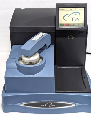
TA Instruments Differential Scanning Calorimeter Q2000

The Differential Scanning Calorimeter (DSC) Q2000 is a highly advanced thermal analysis tool, featuring a temperature range from -90°C to 725°C and exceptional sensitivity for detecting minute heat flow changes. It is designed to precisely measure thermal transitions such as melting, crystallization, and glass transitions, providing detailed insights into material properties. Equipped with high-resolution sensors and intuitive software, the DSC Q2000 offers accurate and reproducible thermal data, making it an essential instrument for material characterization and analysis.

Gel Permeation Chromatography Systems

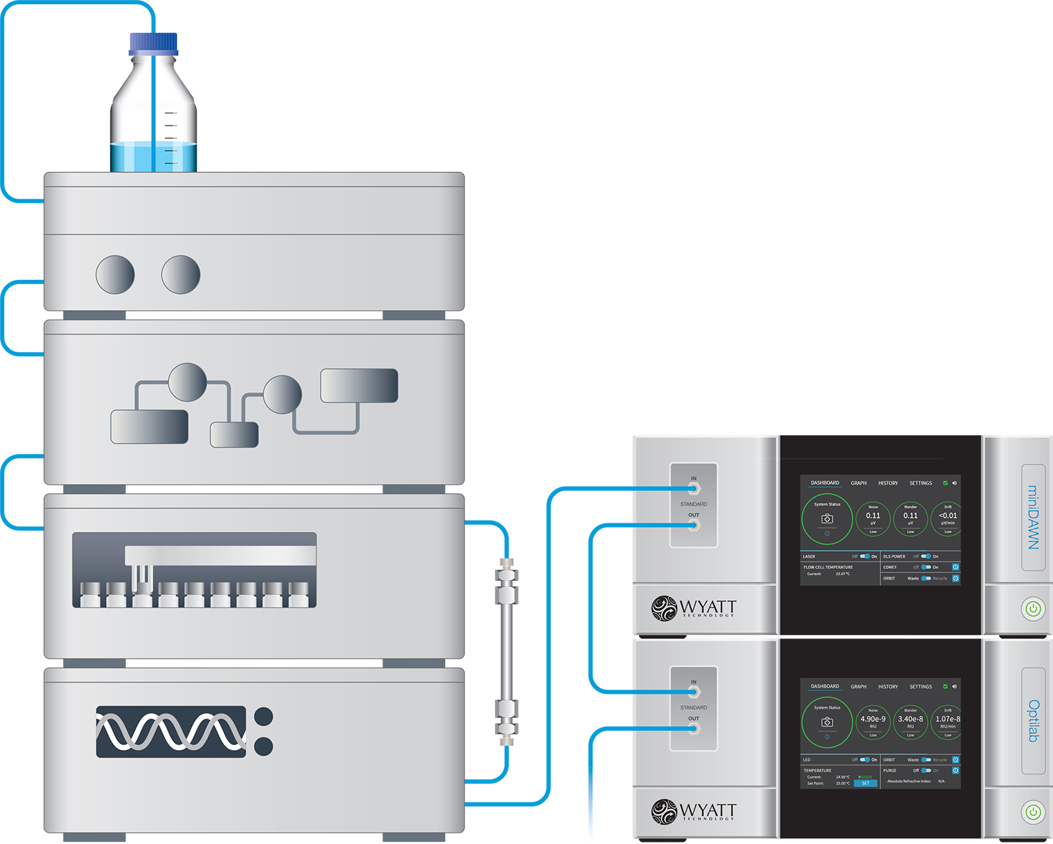
Two Gel Permeation Chromatography (GPC) systems are available for use in the Biomaterials Characterization Suite: One organic and one aqueous system. Both systems utilize Agilent 1260 Infinity II LC Systems with inline Wyatt miniDAWN TREOS light scattering detectors and Opti-TrEX refractive index detectors. Columns can be provided by the user or we have a range of columns available for use.

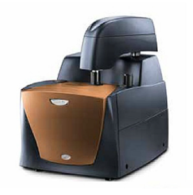
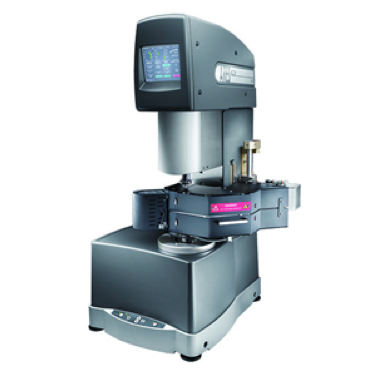
ARES G2 Rheometer

The ARES-G2 Rheometer is a sophisticated tool for detailed rheological analysis, offering broad temperature and viscosity ranges. It accurately measures viscosity, shear stress, and shear rate, and supports advanced tests like oscillatory and transient measurements. Equipped with high-precision torque transducers and versatile measurement geometries, the ARES-G2 provides comprehensive insights into material flow and deformation, making it a valuable asset for both research and industrial use.

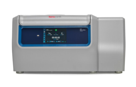
Sorvall X Pro Series Centrifuge

The Sorvall X Pro Series Centrifuge integrates a powerful drive system with an intelligent user interface. This innovative control system optimizes rotor acceleration, resulting in efficient sample separation. Its adaptive control algorithms cater to various experimental scenarios, ensuring safe and reproducible results across diverse sample types and sizes.

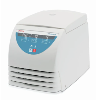
Sorvall Legend Micro 21R Microcentrifuge

The Sorvall Legend Micro 21R Microcentrifuge combines intricate rotor technology with precise motor control for optimal sample separation. The brushless induction drive motor offers high acceleration and deceleration rates, ensuring rapid and consistent sample processing. Its temperature control and safety features enable reproducible results while minimizing sample degradation.

Biomolecular Analysis Suite

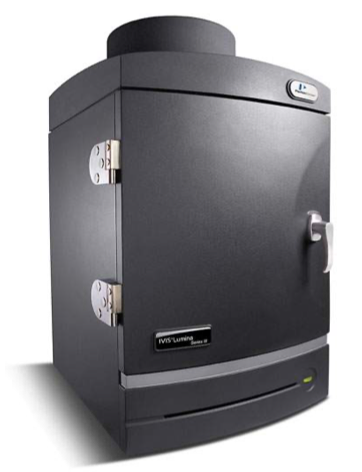
IVIS Lumina III Series

The IVIS Lumina Series III is a state-of-the-art optical imaging system for non-invasive in vivo and in vitro studies. It employs bioluminescence and fluorescence imaging to monitor gene expression, molecular pathways, and cell tracking. With a sensitive CCD camera, it quantifies signals accurately and allows multi-fluorophore imaging. Valuable for preclinical research, it provides insights into disease models, drug effects, and therapeutic responses, reducing the need for invasive sampling.

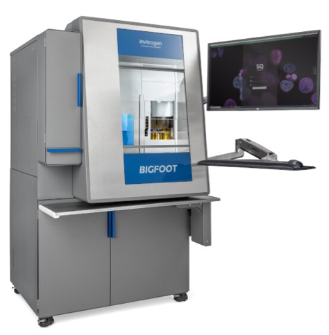
Bigfoot Spectral Cell Sorter

The Bigfoot Spectral Cell Sorter employs cutting-edge spectral cytometry technology, integrating laser excitation and advanced wavelength analysis to segregate cells. This methodology precisely identifies cells through their distinct fluorescent markers, enabling multiparametric characterization. The subsequent droplet-based sorting employs intricate hydrodynamic forces to isolate specific cell populations with exceptional precision, making it invaluable for genomics and immunophenotyping studies.

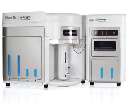
Attune NxT Flow Cytometer

The Attune NxT Flow Cytometer employs innovative hydrodynamic focusing and laser-induced fluorescence excitation techniques. This enables the analysis of cells at the individual level, yielding valuable fluorescence emission and scatter data for detailed cellular attribute assessment. This high-speed cytometer excels in applications such as cell cycle analysis, apoptosis studies, and identification of rare cell subsets, offering in-depth and accurate cellular characterization.

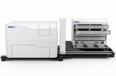
Cytation 10 Confocal Imaging Reader w/ BioSpa Automated Incubator

The Cytation C10 Confocal Imaging Reader combined with the BioSpa 8 Automated Incubator offers enhanced live-cell imaging and analysis. The confocal imaging system provides high-resolution images by eliminating out-of-focus light through laser scanning. Its spectral detection capability enables multiplexed labeling experiments. The BioSpa 8 Incubator maintains optimal conditions for extended live-cell studies. This duo is crucial for observing dynamic cellular processes and interactions in real time.

Cytation 5 Multi-Mode Plate Reader

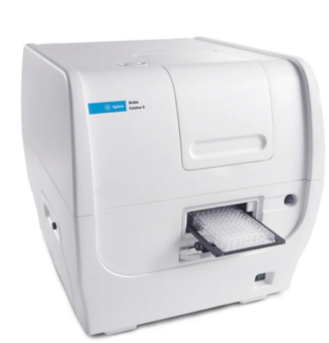
The Cytation 5 Cell Imaging Multi-Mode Reader uniquely integrates advanced microscopy and multi-mode detection capabilities. Its microscopy module captures high-resolution images of multiwell plates, enabling precise cell counting and comprehensive morphological analyses. The multi-mode detection unit combines fluorescence, luminescence, and absorbance measurements in a single platform, facilitating diverse high-throughput cellular assays with unprecedented versatility.

Lionheart LX Automated Microscope

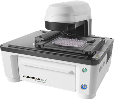
The Lionheart LX Automated Microscope integrates precision optics with motorized stage control and customizable automation protocols. The optical system offers brightfield and fluorescence imaging modalities with adjustable illumination and filters. Combining cutting-edge mechanics and electronics, this system automates imaging processes, minimizing user intervention for live-cell imaging, kinetic assays, and detailed image-based studies.

Seahorse XFe96 Analyzer

The Seahorse XFe96 Analyzer is a progressive instrument used to study cellular metabolism. It employs real-time, label-free analysis in a microplate format to assess mitochondrial function and energy dynamics. By precisely controlling oxygen and pH levels, it monitors oxygen consumption rate (OCR) and extracellular acidification rate (ECAR), unveiling cellular respiration and glycolysis. Chemical probes enable dynamic measurements of metabolic parameters, making it essential for diverse research like cancer biology, immunology, and drug discovery.

iBright CL750 Imaging System

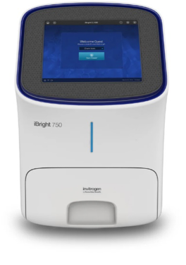
The iBright CL750 Imaging System merges chemiluminescence and fluorescence imaging capabilities with a high-sensitivity camera. This combination offers optimal signal detection and quantification. With precise temperature control and specialized filters, the system delivers high-resolution images critical for accurate protein analysis, western blotting, and gel documentation. Its versatile software enables quantitative analysis and adaptability to various research needs.

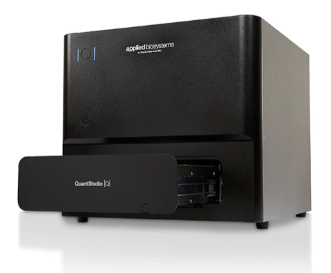
QuantStudio Absolute Q Digital PCR System

The QuantStudio Absolute Q Digital PCR System employs microfluidic arrays and thermal cycling control to divide samples into a multitude of reaction wells. This advanced thermal and fluidic management ensures precise temperature uniformity, enabling digital PCR reactions with high precision. The microfluidic array design guarantees single-molecule partitioning, essential for absolute nucleic acid quantification and rare variant detection.

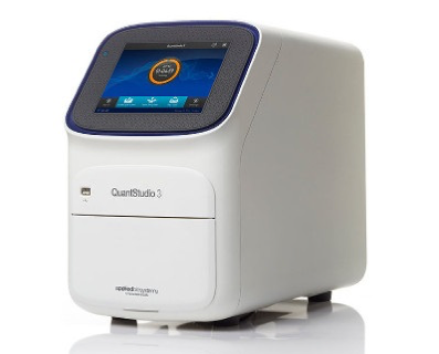
QuantStudio 3 Real-Time PCR System

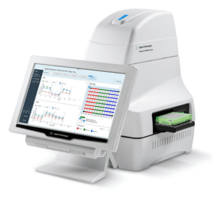
The QuantStudio 3 Real-Time PCR System employs cutting-edge optical technology for fluorescence detection during PCR cycling. The system's advanced optics and thermal management enable real-time monitoring of amplification kinetics. Its software analyzes raw fluorescence data to derive quantitative information. The instrument's rapid cycling capabilities and sophisticated algorithms support various quantitative PCR applications.

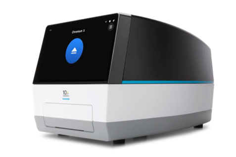
Chromium iX Single Cell System

The Chromium iX Single Cell System leverages a microfluidic chip that encapsulates single cells with barcoded beads. The precisely engineered oil-coated microfluidic channels create aqueous droplets containing individual cells and barcoded beads. This method enables high-throughput, single-cell genomics by capturing cells and barcodes in separate compartments, subsequently enabling multi-omics profiling.

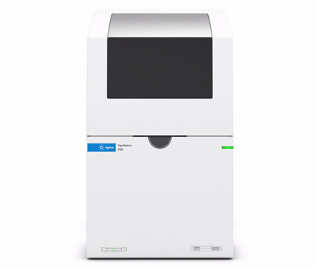
4150 TapeStation System

The 4150 TapeStation System revolutionizes nucleic acid and protein analysis with automated capillary electrophoresis. Its microfluidic chip integrates separation, detection, and quantification. The precisely controlled electric fields enable rapid sample separation in gel-like matrices. The system's software translates migration rates into quality metrics, facilitating automated sample quality assessment, sizing, and quantification.

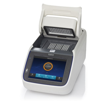
SimpliAmp Thermal Cycler

The SimpliAmp Thermal Cycler utilizes leading-edge technology for accurate and rapid temperature control. The thermoelectric module rapidly alternates between temperature setpoints, enabling precise PCR cycling. Its intuitive interface allows users to design and execute temperature profiles with ease, supporting diverse PCR applications, from routine to complex gene expression studies.

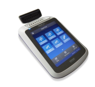
Qubit Flex Fluorometer

The Qubit Flex Fluorometer employs targeted fluorometric assays with precise excitation and emission wavelengths. It selectively measures fluorescent signals from DNA, RNA, and proteins, effectively eliminating background noise. Its proprietary dye-based quantification system ensures high sensitivity and broad dynamic range, offering accurate quantification across a range of concentrations.

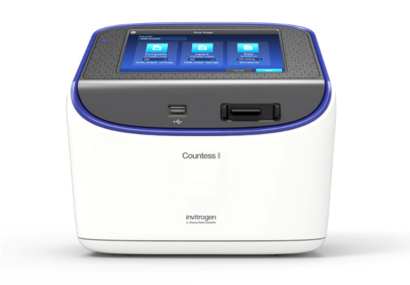
Countess II Automated Cell Counter

The Countess II Automated Cell Counter leverages sophisticated brightfield imaging and proprietary image analysis algorithms. Its integrated optical system captures high-resolution images of cells, and the algorithmic processing swiftly detects and quantifies cells, along with assessing their viability and size distribution. Automation reduces user variability, ensuring consistent and reliable cell culture analysis.