
UNDER CONSTRUCTION
Graphene Core Testing Facility Instrumentation
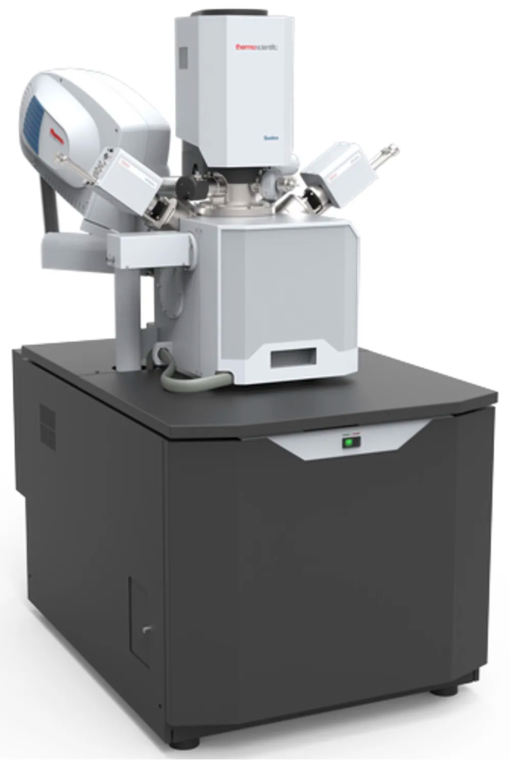

Quattro S Field-Emission Environmental Scanning Transmission Electron Microscope
The Quattro STM is a field emission environmental scanning electron microscope (ESEM) capable of generating and collecting all available information from any type of sample material. it is the most versatile high-resolution, low-vacuum field-emission gun (FEG) SEM with extended low-vacuum capabilities for really challenging samples and dynamic experiments. The Quattro S is the most versatile high-resolution SEM for characterization, prototyping and in situ analysis. This easy to use microscope can provide many research solutions and can be further extended with available options. The Quattro S can freely and simply be switched between three vacuum modes, enabling investigation of conductive, non-conductive and high-vacuum incompatible materials:
- High-vacuum mode (6x10⁻⁴ Pa) for imaging and microanalysis of conductive and/or conventionally prepared specimens
- Low-vacuum mode (10-130 Pa) for imaging and microanalysis of non-conductive specimens without preparation
- ESEM™ mode (10-4000 Pa) for high-vacuum incompatible specimens which are impossible to investigate with traditional EM methods
Features/Specifications:
- Continuously adjustable voltage 200 V to 30 kV (lowest landing energy of 200 V standard and 20 V with beam deceleration mode) and a beam current of up to 200 nA
- Magnification of 20x (at WD = 50 mm) to more than 1,000,000x in single quadrant view of the Quattro S user interface on the standard 24” LCD monitor
- Identical field of view in high- and low-vacuum modes (21 mm at the longest working distance) and 500 µm with standard, axial, gaseous secondary electron (SE) detector
- Pathfinder Alpine energy dispersive X-ray spectroscopy (EDS) System with UltraDry premium detector for analytical chamber SEM with an active area of 60 mm2 and 129 eV energy resolution at Mn k-alpha
- Resolution of 0.8 nm at 30 kV (STEM), 1.0 nm at 30 kV (SE) in high vacuum, 1.3 nm at 30 kV (SE) in low vacuum and ESEM mode, and 3.0 nm at 1 kV (SE)
- Retractable directional back-scattered (DBS) detector
- Retractable STEM 3+ detector
- Solid-state detector integration kit
- 1000°C heating stage to record in-situ morphological sample changes in 5-mm ceramic crucibles
- Cooling/heating stage control kit
- Cooling stage (0-10°)C to image and analyze specimens at relative humidity conditions up to 100% at chamber pressures of 3-10 mbar and possibility of in-situ freeze-drying when the temperature range of the cooling stage is set below 0°C with a minimum of -20°C
- ColorSEM for always-on, live EDS analysis
- In-chamber Nav-Cam for photo-based sample navigation
- Integrated plasma cleaner for chamber cleaning and/or a mild specimen surface cleaning for the removal of hydrocarbon contaminants when operating at low kV
- Multi-purpose SEM holder with labeled positions and unique stage mounting, allowing simultaneous loading of 18 standard samples (ø 12 mm), three 45˚ pre-tilted samples, and two row bars (vertical, and 52˚ pre-tilted)
- Maps 3 software for SEM/SDB
Brochure: Quattro S SEM
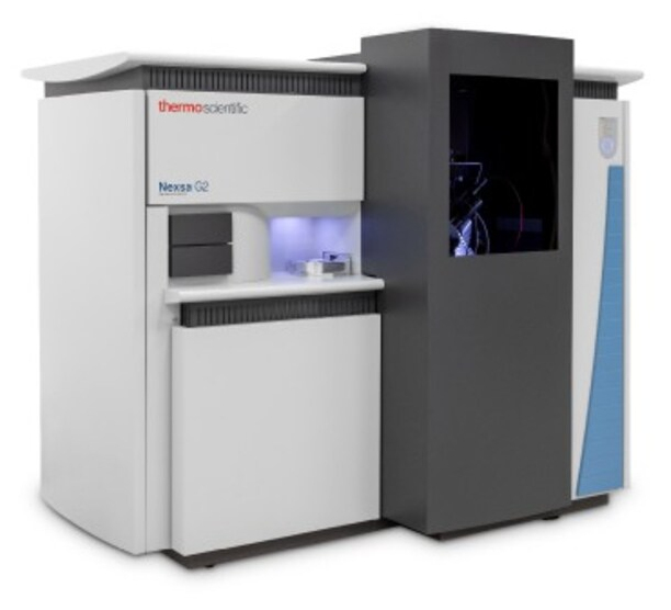

Nexsa G2 Surface Analysis (X-ray Photoelectron Spectroscopy) System
The NexsaTM G2 XPS System offers fully automated multi-technique analysis, for high throughput, research grade results. A high specification micro-focused Al Kα X-ray source is complemented by patented features such as co-axial, dual-beam charge compensation for insulator analysis, and reflex optical viewing to allow rapid identification and analysis of small features. The XPS SnapMap feature further enhances analysis by enabling rapid mapping of sample chemistry over small areas, to identify and detect subtle differences which may not be detectable otherwise. The Nexsa G2 system is equipped with ion scattering spectroscopy (ISS), UV photoelectron spectroscopy (UPS), reflected electron energy loss spectroscopy (REELS), and Raman spectroscopy (RS), integrated onto the same platform, which allows users to conduct true correlative analysis. This capability unlocks the potential for analysis to drive advances in microelectronics, ultra-thin films, nanotechnology development and many other applications.
Features/Specifications:
- Double-focusing hemispherical electron energy analyzer with multi-element input lens, 125 mm mean radius, full 180° hemispherical analyzer, and 128 channel position sensitive detector
- Microfocused 0.25 m Rowland circle monochromated x-ray source,12 keV nominal operating voltage electron source, motorized, water-cooled aluminum-coated anode with >20 operating positions, and single toroidal quartz crystal
- User selectable x-ray spot sizes in 5 µm steps in the range of 10-400 µm and 120 W maximum power (400 µm X-ray spot)
- XPS SnapMap rapid XPS imaging capability
- 4-axis specimen stage with two multi-specimen mounting plates and maximum specimen dimensions of 60x60x20 mm, one mounting plate for powder samples, one mounting plate for fiber samples, one set of three rotation holders (maximum specimen dimensions 30 mm diameter, 15 mm thick), and one mounting plate for use in combination with a rotation holder
- Tilt sample holder for angle-dependent XPS in the range of +/-90° with respect to the surface normal and maximum specimen dimension of 26x5x5 mm
- Vacuum transfer module to allow samples with a maximum specimen thickness of 9 mm to be transferred under vacuum into the system
- Automated specimen transfer mechanism controlled from the Avantage software
- Avantage v6 Software with full control of all aspects of XPS data acquisition (including spectra, SAXPS, line scans, maps, depth profiles)
- Bipolar analyzer electronics (XPS+ISS)to allow both negative (electrons) and positive (ions) energy selection with the kinetic energy range of 0–1500 eV, minimum energy step size of 3 meV, and pass energy of 1-400 eV (continuously selectable)
- iXR Raman spectrometer with optical integration to the analysis chamber and alignment to the XPS analysis position, 532±1 nm laser (10.0 mW laser power resolution of 0.1 mW, spot size at sample of <15 µm, 25 and 50 µm pinhole apertures, 25 and 50 µm slit apertures, spectral resolution better than 5.0 cm-1 FWHM, spectral dispersion of 2 cm-1/CCD pixel element, upper cutoff of 3400 cm-1, and lower cutoff of 50 cm-1
- Reflected electron energy loss spectroscopy (REELS) and charge compensation combined low-energy electron/ion flood source for charge neutralization and REELS analysis with electron beam energy of 0-10 eV for charge compensation, electron beam energy of 0-1000 eV for REELS, and electron beam emission of up to 250 µA
- Monatomic and gas (helium or argon) cluster ion source (MAGCIS) with automated beam alignment and focusing, differentially pumped electron impact source with dual electrostatic lenses and floating flight tube, beam energy in monatomic mode of 500-4000 eV, beam energy in cluster mode of 2000-80000 eV, and cluster size range of 75-2000 atoms
Brochure: Nexsa G2 XPS
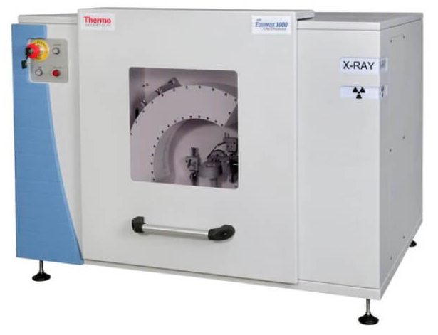

ARL EQUINOX 100 X-ray Diffractometer
ARL EQUINOX 100 is a diffractometer with fully integrated water cooling system, suitable for qualitative and quantitative analysis, phase identification, structure determination, crystallites orientation, thin film, and dynamic studies. Thanks to its detection mode, real time measurements without goniometric movement (simplified goniometer without motorization) are possible on a 110°/2θ angular range.
Features/Specifications:
Brochure: ARL EQUINOX 100 XRD
![]()
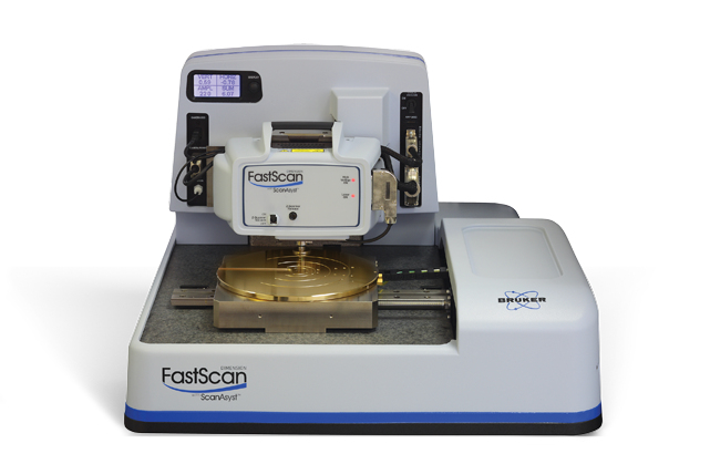
Dimension FastScan Atomic Force Microscope
Brochure: Dimension FastScan AFM
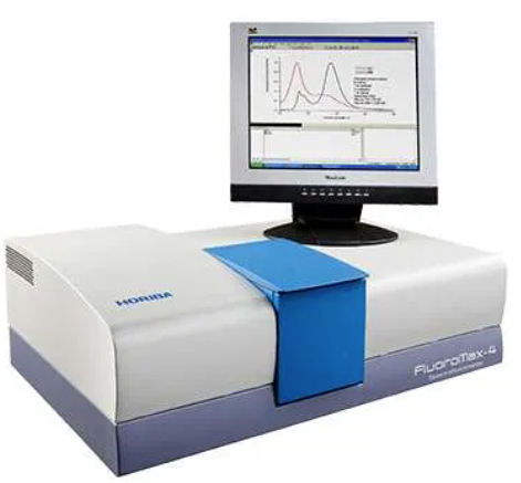

Scientific FluoroMaxTM Plus Spectrofluorometer
- Fluoromax Plus all-reflective-optics research spectrofluorometer with 10,000:1 water Raman specification (FSD method)
- Continuous 150W ozone-free xenon source delivering excitation light from 230nm to the NIR
- DSS-IGA020L liquid nitrogen cooled InGaAs detector (800-1700nm at RT, 800-1550nm at LN2 temperature)
- Mounted into the all-reflective-optic 1427C-AU housing
- Rapid Peltier temperature controlled single sample holder, no circulating water is required for normal operation from 0 to +110°C
- QuantaPhi-2 Integrating sphere for direct PLQY sample measurements
- 121 mm internal diameter Spectralon® integrating sphere with reflectivity from 250-2500nm
Brochure: FluoroMax Plus SFM
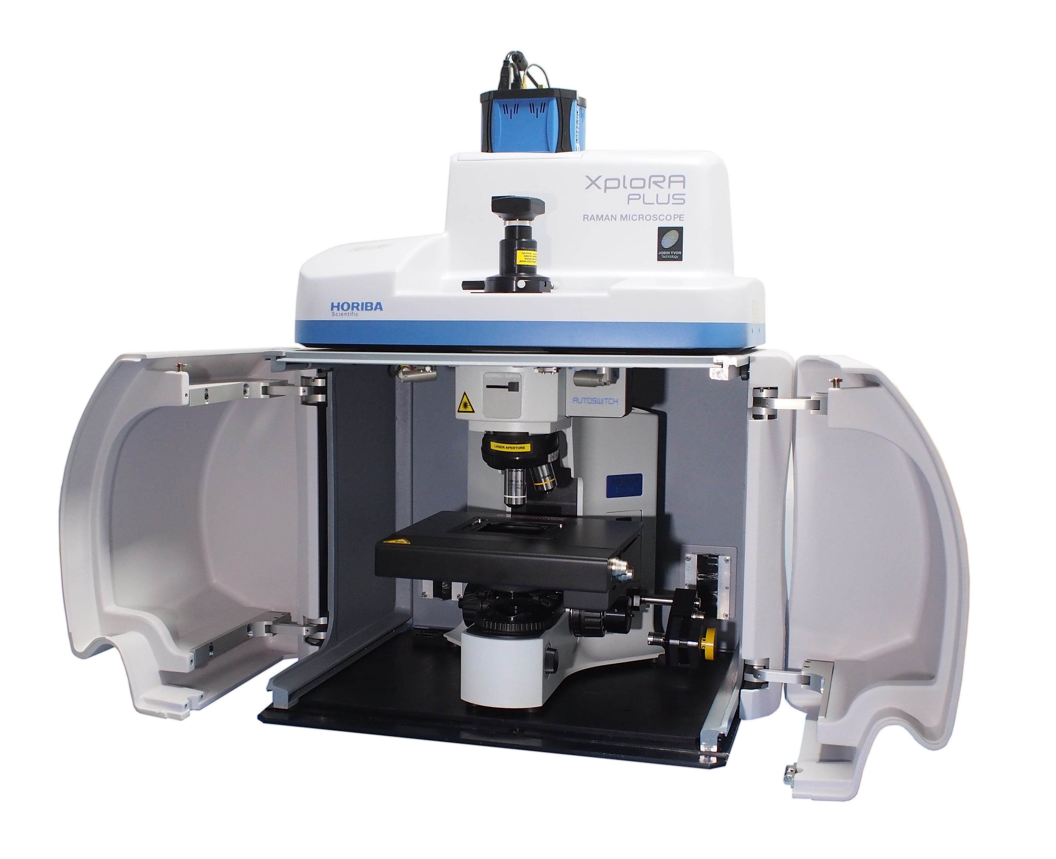

XploRATM PLUS MicroRaman Spectrometer Confocal Raman Microscope
Brochure: XploRA PLUS RM
General Facility Instrumentation

Nicolet iS50 Fourier-Transform Infrared Spectrometer
Brochure: Nicolet iS50 FTIR
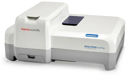

EVOLUTION One Plus UV-Visible Spectrophotometer
Brochure: EVOLUTION One Plus UV-Vis Spectrophotometer
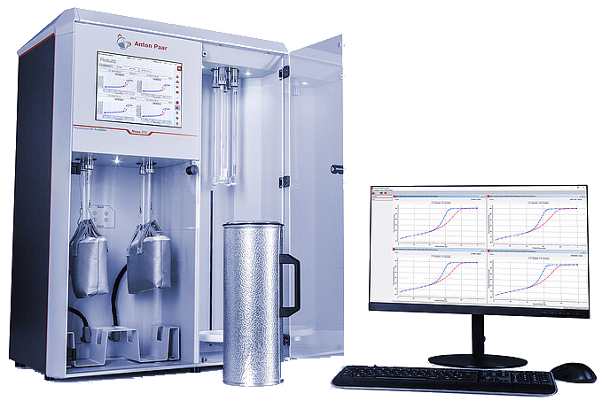

NOVATM 600 Surface Area and Pore Size Analyzer
Brochure: NOVA 600 Surface Area and Pore Size Analyzer
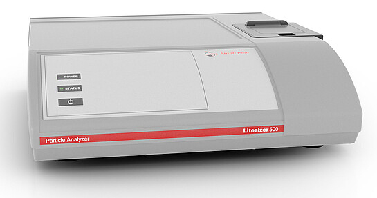

LitesizerTM 500 Particle Size Analyzer
Brochure: Litesizer 500 Particle Size Analyzer
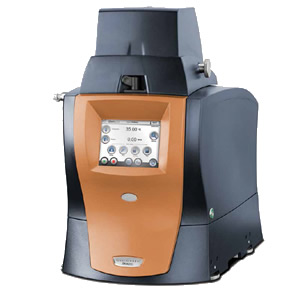

Discovery DMA 850 Dynamic Mechanical Analyzer
Brochure: Discovery DMA 850
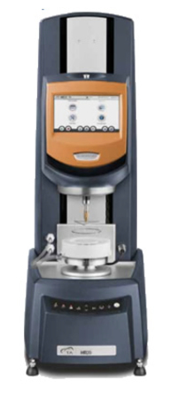

Discovery HR 20 Hybrid Rheometer
Brochure: Discovery HR 20 Hybrid Rheometer
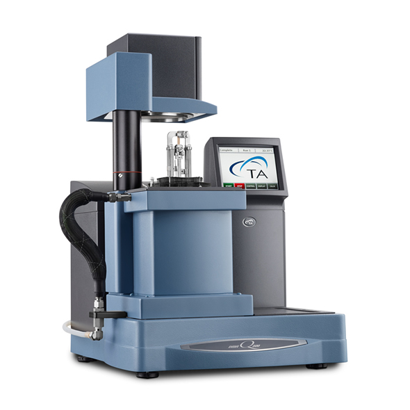

DMA Q800 Dynamic Mechanical Analyzer
Brochure: DMA Q800 Dynamic Mechanical Analyzer

TGA Q500 Thermogravimetric Analyzer
Brochure: TGA Q500 Thermogravimetric Analyzer

Thermal Analysis System TGA/DSC 3+
Brochure: TGA/DSC 3+ Thermogravimetric Analyzer and Differential Scanning Calorimeter
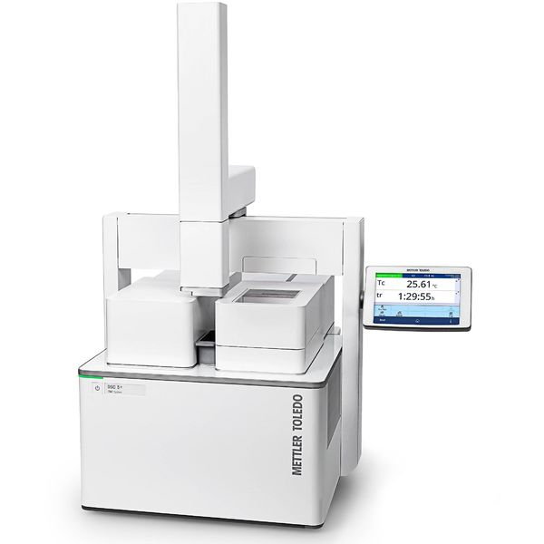

Thermal Analysis System DSC 5+
Brochure: DSC 5+ Differential Scanning Calorimeter
Materials Synthesis and Processing Equipment

Nano Thermal Chemical Vapor Deposition (TCVD) System for MoS2, WS2, and Graphene Nanomaterials
Brochure:
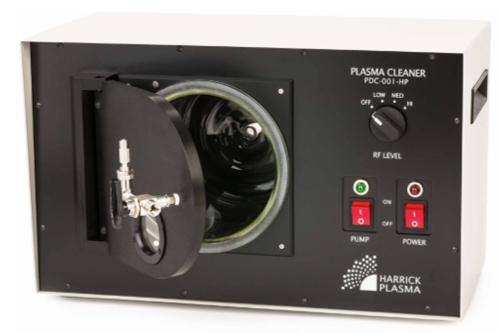

High Power Expanded Plasma Cleaner
Brochure: High Power Expanded Plasma Cleaner
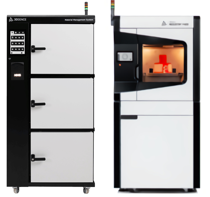
![]()
INDUSTRY F421 High-Performance 3D Printer with Material Management System
Brochures: INDUSTRY F421 High-Performance 3D Printer and Material Management System
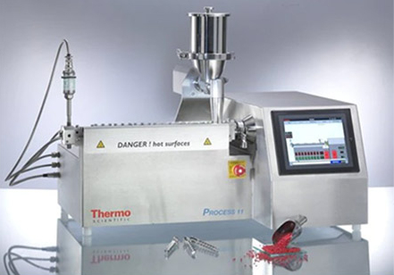

Process 11 Parallel Twin-Screw Extruder
Brochure: Process 11 Parallel Twin-Screw Extruder
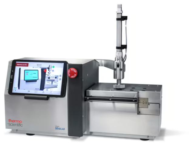

HAAKE™ MiniLab 3 Micro Compounder
Brochure: HAAKE MiniLab 3 Micro Compounder

Micro-18 Modular Twin-Screw Extruder

